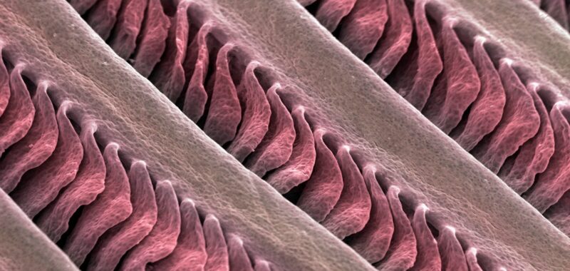
Evolution is a master recycler. It often uses old structures (or ancient genes) for new jobs. The mammalian ear is a perfect example. Over the eons, the jawbones of our fish ancestors became three separate small bones that transmit sound waves from the eardrum to the inner ear.
Now a new study shows that there was another hand-me-down from fish to mammals. It turns out that the flexible cartilage of fish gills bears a close correspondence to the cartilage in the mammalian outer ear, the visible part of the ear. To be sure, flexible cartilage structures take on different jobs in fish and mammals: the gill structures enable fish to breathe while the cartilage in mammals’ outer ears captures sound. But the underlying gene network that builds these structures shares a common history.
To be clear, the gill structures did not morph into the mammalian outer ear. Rather, as the first vertebrates emerged on land and dispensed with gills, the underlying network of genes that formed the gill cartilage was able to build something new. “That’s one of the amazements of life and evolution,” says Abigail Tucker, a professor of development and evolution at King’s College London, who was not involved in the study. “The regulatory network was still there and therefore could be co-opted and used again, this time to make an external ear structure rather than a gill.”
On supporting science journalism
If you’re enjoying this article, consider supporting our award-winning journalism by subscribing. By purchasing a subscription you are helping to ensure the future of impactful stories about the discoveries and ideas shaping our world today.
This recycling of the same gene network provided the foundation for subsequent evolutionary innovations. The cartilage in the mammalian outer ear evolved further into a variety of shapes, such as those of the large, sensitive ears of echolocating bats, the alert, pointed ears of a cat or the floppy ears of an elephant—which are each attuned to the sounds that are important to that animal. In some mammals, the ear cartilage has even become further modified, filled with specialized cartilage cells that contain large fat droplets that researchers hypothesize give the cartilage unique structural and acoustic properties.
“We think there is an ancestral program to make cartilage-filled gills in the head that has moved in position during evolution to become more closely associated with the ear, much as the ancestral fish jawbones moved into the middle ear,” explains Gage Crump, a stem cell biologist at the University of Southern California and senior author of the new study, which was published in Nature. “The program to grow out a cartilaginous structure in this general area of the head is deeply conserved, but the exact position, the full repertoire of expressed genes, and hence the types of cells and their functions have changed quite a lot.”
Crump’s team, which uses zebrafish as a model organism for its research, had long been interested in the development of the vertebrate face. When building an atlas of all the different cell types in the zebrafish face, the researchers noticed two types of cartilage, one that was expected and one that they hadn’t noticed before. This unexpected form was a rod of elastic cartilage that supported the gills’ fingerlike projections. That cartilage was similar to the type found in mammals’ outer ear.
The researchers observed that gene activity in the human outer ear cartilage was similar to that in the elastic cartilage in the fish gills. But many genes are active in unrelated organs. To see if the structures shared an evolutionary history, the researchers focused on the enhancers—the sequences of DNA that drive the activity of their target genes in a specific tissue. They identified six key enhancers that were essential for development of cartilage in the human outer ear but not in the nose. The researchers reasoned that if gene activity in fish gills and mammalian ear cartilage was initiated by similar enhancers, then these structures would be likely to share the same evolutionary origin.
This approach focusing on the enhancers “is very inspiring and really very intelligent, very astute,” says Licia Selleri, a stem cell and developmental biologist at the University of California, San Francisco, who was not involved with the research. “This could reveal whether the new structures arose from the utilization of an ancestral developmental program or if they appeared de novo.”
To investigate the fish-gill-ear question, the researchers, led by Crump’s then graduate student Mathi Thiruppathy, did some clever genetic transfer experiments. First, they put the six human outer ear enhancers that control genes into the genome of zebrafish, and they used a fluorescent reporter gene that lit up to identify the location in the body where the enhancers’ usual targets would normally be activated. Strikingly, the human ear cartilage enhancers only drove the activity of green fluorescent protein in the zebrafish gills, suggesting that whatever is controlling gene expression is very similar between the gills and outer ear, Crump says.
Then the team did another experiment: it put the key enhancers that were active in the zebrafish gills into the mouse genome. There, the researchers observed that the fish DNA elements now activated the green fluorescent protein in the developing transgenic mice’s outer ear, reinforcing the idea that the same underlying gene network was being used to build cartilage in the gills and the ear.
“The bit that makes it more interesting than just reusing the same molecular toolkit is the fact that it also reuses the regulatory elements [enhancers] that control the expression of those genes,” so the regulatory elements that drive expression of genes for cartilage in the gills drive the expression of cartilage genes in the mammalian ear, Tucker says. “So it’s got an extra level of using the system that was there before.”
Next the researchers sought to identify which key genes were under the influence of these enhancers. One gene family that stood out was DLX, which is related to a gene identified in the fruit fly as distal-less that is important for insect limb development. The researchers found that the same enhancers for the vertebrate DLX genes turned up in animals from zebrafish to humans over more than 400 million years of evolution. That is why the enhancers were able to be swapped into the genetically engineered fish and mice.
To see just how old these enhancers were, the researchers looked at horseshoe crabs, invertebrates that also breathe with gills. They discovered that the same distal-less gene that is related to the DLX gene is also involved in making the gills of horseshoe crabs. And by plugging the DNA control element from horseshoe crabs into the zebrafish genome, it was possible to activate the fluorescent molecule in the zebrafish gills. This suggests that the genetic machinery that makes the mammalian outer ear predates the evolution of vertebrates; it may date back hundreds of millions of years to some of the first marine invertebrates that had gill-like projections. When fishes, the first vertebrates, evolved, the gene network that builds gill cartilage from those invertebrates was recycled to make fish gills, even as fish evolved a new type of bony skeleton.
“We think that the elastic cartilage in our outer ears may be the last remnant of invertebrate cartilage,” Crump speculates.
To understand what occurred along the vertebrate evolutionary tree between fish and mammals, the researchers looked at the activity of those same enhancers in frogs and lizards. In frog tadpoles, the human outer ear enhancers activated the fluorescent protein in the tadpole gills. In anole lizards, which do not have gills or an outer ear, the human outer ear enhancers activated the fluorescent protein in the animals’ ear canal, which also has an elastic cartilage similar to that in the fish and tadpole gills. This suggests the gene network that makes the elastic cartilage in the fish gills became active in reptiles’ ear canal first and then in mammals’ outer ear.
“So what we imagine is: between the amphibians and reptiles, there’s a shift from the gills to the ear canal that then, in mammals, became massively elaborated to form the outer ear,” Crump says.
Over evolutionary time, the outer ear cartilage in mammals continued to evolve not only in shape but also in internal composition. Cell biologist Maksim Plikus and his team at the University of California, Irvine, recently described cartilage cells in the ears of small mammals—mice, shrews, bats and rats, among others—that are sort of a cross between cartilage cells and fat cells. These cells, which are filled with droplets of fat, form a Bubble Wrap–like tissue called lipocartilage. Although this tissue was first discovered in 1854 by German histologist Franz von Leydig, it had largely been forgotten about until now. Plikus’s team hypothesizes that lipocartilage has unique acoustic properties, such as the ability to increase the propagation of sound waves, which may be an adaptation for mammalian hearing.
“While there is indeed a program that is present in invertebrates and then is reutilized in fish, and in mammals to make the outer ear, there are also innovations that appear in mammals,” says Selleri, who wrote a perspective article in Science about the lipocartilage study. “One of these innovations is the presence of cartilage with fat.”
“[Lipocartilage can] can use vacuoles—lipid droplets—for a completely different purpose than what is normally believed they play,” Plikus says. While the main purpose of lipid droplets in fat cells is to store energy, in lipocartilage, “these lipid droplets primarily play a structural and biomechanical role, so they no longer contribute to a metabolic function,” Plikus says.
Postdoctoral researcher Raul Ramos led the study, published in Science. The researchers showed that in mice, the fat vacuoles are unchanging in response to metabolic state: they do not increase in size when a mouse is overfed, and the fat droplets are not used for energy when the animal is starved. The team further showed that the droplets are madeusing a very specific metabolic pathway that transforms sugars into fat—and this controlled metabolic pathway allows the animal’s body to regulate the exact size and spacing of the lipid droplets.
That, in turn, enabled the evolution of ear structures with acoustic properties suited to the needs of various types of animals—bats’ large, ridged ears, for instance, are so sensitive they can detect the flap of the wings of a tiny insect.

